How a General Dentist Detects Dental Problems Early
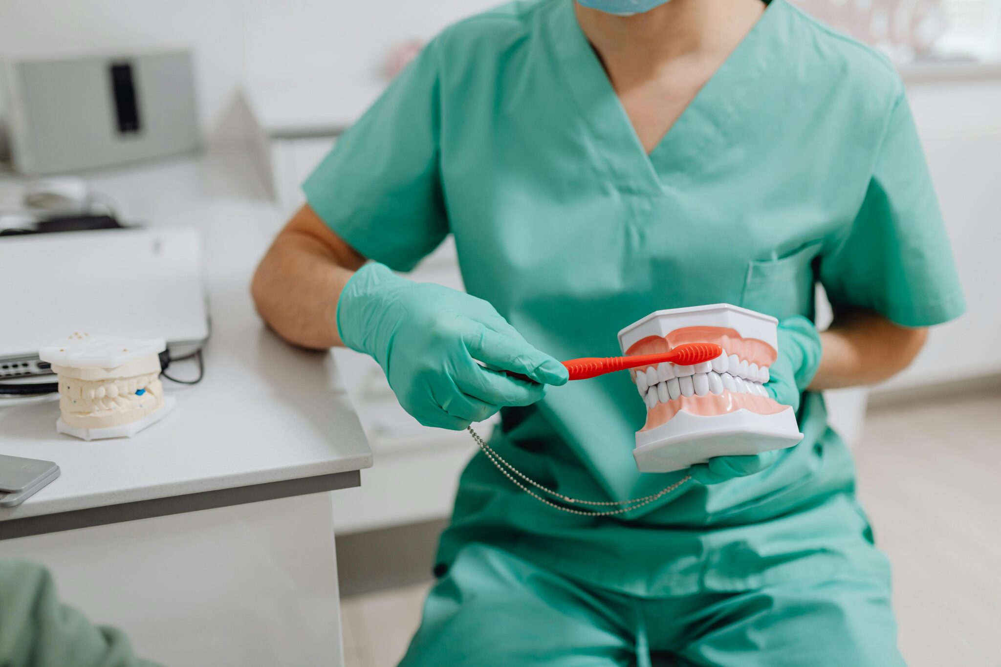
Most dental problems do not begin with dramatic pain. They start quietly, sometimes invisibly, with a small change in enamel, a slight swelling in the gums, a shift in bite pressure, or a shadow on an X-ray that means little to a patient and quite a lot to a trained eye. That quiet beginning is exactly why early detection matters so much in general dentistry.
A good general dentist is not simply waiting for cavities to appear or for a tooth to crack. The real work is more observant than that. It involves pattern recognition, careful listening, comparison over time, and a surprisingly detailed understanding of how oral tissues behave when something is just beginning to go wrong. Patients often think the appointment is about cleaning and checking for “anything obvious.” In practice, the visit is much more nuanced.
Early detection can mean the difference between a small filling and a root canal, between reversing gum inflammation and losing bone support, between monitoring a harmless area and catching a lesion that needs urgent referral. The value is not only clinical. It is financial, practical, and personal. Small problems are usually easier to treat, cheaper to manage, and less disruptive to daily life.
The exam starts before instruments touch the teeth
Experienced dentists begin gathering information before the formal examination starts. The way a patient speaks, opens the mouth, swallows, or describes discomfort can offer useful clues. A person who says, “It only hurts when I bite on something hard,” is describing a different problem from someone who says, “Cold water sets it off for a minute,” and different again from someone with pressure, swelling, and a dull ache that wakes them at night.
Even body language can matter. Patients with jaw joint strain often rub the side of the face without realizing it. People who grind their teeth may present with tight jaw muscles, chipped front teeth, or a complaint that their teeth feel “shorter” or sensitive in the morning. Someone embarrassed about bleeding gums may mention it casually while discussing mouthwash, yet that small comment can lead to the discovery of active periodontal disease.
Medical history also sharpens what the general dentist looks for. Dry mouth in a patient taking several medications raises the risk of rapid decay, especially along the roots. Diabetes can change how gum disease behaves. Acid reflux, eating disorders, and certain diets can leave distinct erosion patterns on tooth surfaces. Pregnancy can temporarily alter gum tissue response. None of these details guarantee a diagnosis, but they guide attention.
Visual clues are often subtle, not dramatic
The popular image of dental disease is a black hole in a tooth. Real life is often less obvious. Early enamel decay may look like a chalky white area near the gumline or between teeth. Demineralization can show up as a dull spot on a tooth that should be glossy. Gum disease may begin as puffiness, color change, or bleeding during gentle probing, long before teeth feel loose.
General dentists are trained to notice small deviations from normal anatomy. They look at symmetry, contour, color, texture, and cleanliness. They compare one side of the mouth with the other. They check whether an old filling has a margin that no longer blends smoothly with the tooth. They notice a hairline crack that catches light differently. They look for flattening on chewing surfaces, shiny wear facets from grinding, and recession that exposes root surfaces vulnerable to sensitivity and decay.
Soft tissues matter just as much as teeth. The cheeks, tongue, floor of the mouth, palate, and lips can reveal ulcers, frictional changes, fungal infections, salivary gland issues, or lesions that need monitoring or biopsy. Many of these areas cause no pain at first. A patient can feel perfectly fine and still have an abnormal spot that deserves a second look.
That is one of the less appreciated parts of a routine dental visit. The examination is not only about the structures people can see in the mirror. It is also about the places they cannot easily inspect and the changes they would not know how to interpret.
The explorer matters less than judgment
Patients often remember the old-fashioned “poke around” with a metal instrument and assume that is how decay is found. Modern dentistry relies much less on forceful probing than on judgment, lighting, magnification, radiographs, dryness, and a careful understanding of risk.
A sticky feeling in a groove does not always mean a cavity, and pushing hard into suspicious enamel can actually damage an early lesion. Many dentists today prefer to dry the tooth thoroughly and inspect the surface visually. A dry field can reveal texture and opacity changes that are hidden when saliva is present. Good overhead light and magnification can make the difference between spotting an early problem and missing it.
This is where experience counts. Two dark grooves on molars may look equally suspicious to a patient, yet one may be harmless staining while the other reflects active decay under weakened enamel. The distinction often depends on subtle findings, the patient’s cavity history, recent X-rays, fluoride exposure, and whether the area has changed since the last visit.
X-rays show what eyes cannot
Some of the most important dental problems are not visible during a mirror exam. Cavities between teeth, bone loss around roots, infections at the tip of a root, impacted teeth, failing restorations under crowns, and certain cysts or abnormalities may only become apparent on radiographs.
X-rays are not taken on a rigid schedule for every person. A thoughtful general dentist adjusts frequency based on risk. A teenager with multiple recent cavities may need bitewing radiographs more often than an adult with excellent home care and a long history of stable exams. Someone with extensive restorative work, dry mouth, or gum disease may also need closer imaging follow-up.
Interpreting radiographs is not a matter of spotting a dark area and declaring a diagnosis. Radiographic images require context. A shadow may represent decay, overlap, anatomy, or a technical artifact. Bone levels need to be judged against previous images, pocket measurements, and clinical appearance. A small area near a root tip can mean active infection, old scar tissue, or a healing change after prior treatment. The picture matters, but the picture alone is rarely the whole story.
One practical example appears with decay between molars. Patients are often surprised to hear they have a cavity when the tooth looks intact from above and feels normal. Yet once decay starts between teeth, it can progress a fair distance before becoming visible to the naked eye. Bitewing X-rays are especially useful for catching these lesions early, before they undermine a large portion of the tooth.
Gum measurements tell a story over time
Periodontal disease is one of the clearest examples of why routine exams matter. Many patients do not feel it developing. Bleeding while brushing may seem minor. Mild bad breath may be blamed on lunch or dry mouth. Teeth can remain comfortable even as bone support gradually decreases.
That is why periodontal charting is so important. The general dentist or hygienist measures the depth of the space between tooth and gum at multiple points around each tooth. A deeper reading does not automatically mean severe disease, but patterns matter. Bleeding, recession, tartar accumulation, mobility, bone levels on X-rays, and changes from previous visits all help define whether the issue is simple gingivitis, early periodontitis, or more advanced disease.
A six-millimeter pocket around one molar is different from generalized four-millimeter pockets with bleeding throughout the mouth. Localized inflammation around a poorly fitting crown requires one kind of response. Widespread bone loss in a smoker with diabetes requires another. This is where early detection is especially valuable, because gum disease can often be slowed or stabilized far more effectively when caught before major attachment loss has occurred.
I have seen patients genuinely shocked when shown side-by-side radiographs from several years apart. Because the change happened gradually and painlessly, they had no sense that anything serious was unfolding. Once they see the pattern, the reason for treatment becomes much clearer.
Old dental work often gives the first warning
Teeth that have already been treated deserve close attention. Fillings, crowns, bridges, and root canal treated teeth are not “finished” forever. Margins can leak, materials can wear, teeth can crack next to restorations, and decay can recur in places that are hard to clean.
A crown may look acceptable to a patient and still show a slightly open margin under magnification. A composite filling may stain harmlessly at the edge, or the stain may trace a breakdown point where bacteria can enter. A root canal treated tooth may develop tenderness to biting because of a vertical crack, not because the root canal itself has failed.
General dentists spend a lot of time comparing current findings with earlier records. A tiny change around an old filling may not trigger immediate treatment if it is stable and low risk. On the other hand, that same finding in a high-risk patient with active decay elsewhere may justify earlier intervention. Detecting problems early is not just about finding disease, it is also about deciding when watchful monitoring is smarter than drilling and when delay is likely to make matters worse.
Bite patterns reveal stress before pain appears
Teeth do not only suffer from bacteria. Mechanical forces can create their own slow-motion damage. Clenching, grinding, uneven bite contacts, missing teeth, and poorly distributed chewing forces may lead to fractures, sensitivity, muscle pain, gum recession, or joint discomfort.
These issues often announce themselves through patterns rather than one dramatic symptom. A general dentist may see worn incisal edges, tiny craze lines, broken cusps on molars, notching near the gumline, or repeated failure of fillings on specific teeth. None of those signs should be read in isolation. Together, they can reveal excessive bite stress long before a patient experiences a major crack.
This is especially common in busy adults who clench during work or sleep. They may report headaches, neck tightness, or a sense that their teeth “touch too much” in the morning. Sometimes they come in because a corner of a tooth chipped off while eating toast, only to learn that the real issue has been building for years. Catching those wear patterns early can lead to a night guard, bite adjustment in select cases, or restorative planning that prevents a much bigger problem later.
Saliva, plaque, and habits change the risk picture
Not every patient has the same likelihood of developing dental disease. Early detection improves when the dentist understands risk, because risk determines where to look hardest and how often to recheck.
Saliva is one of the best natural defenses in the mouth. When it is reduced by medication, autoimmune disease, cancer therapy, or chronic dehydration, cavities can accelerate quickly. These are not always the classic pits and grooves on molars. Root surfaces may decay near the gumline, front teeth may become vulnerable, and changes can happen in months rather than years.
Home habits matter too. A patient who sips sweetened coffee all morning https://www.google.com/maps?cid=11867611376950550291 presents a different risk pattern from one who drinks it with breakfast and finishes it quickly. Frequent sports drinks, nighttime snacking, poor flossing access around crowded teeth, and inconsistent fluoride use all leave traces in the mouth. The dentist is not only looking at disease. They are reading the conditions that allow disease to start.
Sometimes the earliest sign is a cluster rather than a single lesion. A few chalky areas near orthodontic brackets, repeated small cavities along the gumline, or inflammation concentrated around lower front teeth can all point to habits that need attention before more treatment is needed.
Technology helps, but it does not replace clinical sense
Modern dental offices may use digital radiographs, intraoral cameras, laser fluorescence devices, transillumination, periodontal software, and other tools to support diagnosis. These can be very useful, especially for documenting change and communicating findings to patients. Seeing a magnified crack or an inflamed gum margin on a monitor often helps patients understand what words alone do not convey.
Still, technology is only part of the process. Devices can produce false positives. Images can be misread. A suspicious signal may prompt closer inspection, but it should not automatically dictate treatment. Strong diagnostic dentistry still depends on correlating tools with symptoms, visual findings, tactile information, and history.
The best general dentist does not chase every shadow or every reading from a gadget. They weigh evidence. They ask whether the area is active, stable, restorable, urgent, or simply worth watching. That restraint is part of early detection too. Good care is not just finding more things. It is knowing which findings matter now.
Why follow-up intervals are never one-size-fits-all
Patients often ask how often they really need checkups. The six-month model is common and reasonable for many people, but it is not a law of biology. Some patients benefit from more frequent reviews, while others with low risk and consistently stable oral health may be fine with a longer interval for certain types of assessments, depending on the dentist’s judgment and local standards of care.
A patient with active periodontal treatment may need shorter recall visits. Someone prone to heavy tartar buildup can deteriorate quickly if appointments are spaced too far apart. A person with a history of rapid decay after starting dry-mouth medications may need close monitoring for a year or two. By contrast, a patient with decades of excellent stability may show little change over time.
What matters is not loyalty to a calendar, but the likelihood that a meaningful problem could develop unnoticed between visits. Early detection is strongest when the timing fits the individual.
The patient’s role is larger than many realize
A dentist can only compare what they see today with what they knew yesterday. Patients make that comparison more accurate when they report changes clearly and early. A little sensitivity, food trapping between two teeth, a new rough edge, occasional bleeding in one area, or a sore that has not healed after two weeks can all be worth mentioning. None of these symptoms always signal a serious problem, but they are useful pieces of the diagnostic picture.
It also helps when patients understand that “no pain” does not equal “no disease.” Some of the costliest problems in dentistry remain silent until they are advanced. That includes decay under old restorations, chronic gum disease, cracked teeth, and certain infections. By the time pain appears, treatment is often more invasive.
Patients sometimes worry that regular exams are designed to “find something wrong.” In a well-run practice, the opposite is true. The goal is to keep small findings small, to monitor uncertain areas honestly, and to avoid overtreatment as much as undertreatment. That balance is what makes clinical judgment so important.
What early detection looks like in real practice
In day-to-day dentistry, early detection rarely feels dramatic. It looks like noticing that a contact between two teeth is starting to trap floss. It looks like comparing this year’s bitewing X-rays with the last set and seeing a lesion just beginning to move into dentin. It looks like measuring a gum pocket that was three millimeters last year and is five today with bleeding. It looks like observing that a tooth with a large filling now shows a faint crack line and tenderness on release when biting.
Each of those moments can change the trajectory of care. A small interproximal cavity may need a conservative restoration instead of a crown later. Early periodontal therapy may preserve supporting bone. A protective night guard may save a molar from splitting. A suspicious tissue change may be referred and evaluated before it becomes far more difficult to manage.
That is the central value a general dentist brings to preventive care. The appointment is not just a search for obvious decay. It is a careful review of hard tissue, soft tissue, function, habits, risk, and time-based change. Done well, it turns routine visits into a form of surveillance that protects both oral health and overall well-being.
When people say they want to “stay ahead” of dental problems, this is what staying ahead actually means. It means catching the whisper before it becomes a crisis.
Smyle Dental Newhall
Address: 23754 Newhall Ave, Santa Clarita, CA 91321
Phone number: +16612559200
FAQ About General dentist
What does it mean by general dentist?
A general dentist is your primary dental care provider. They focus on the overall prevention, diagnosis, and treatment of your daily oral health needs. Think of them as your primary care doctor, but for your teeth and gums.
What is the difference between a dentist and a general dentist?
A dentist is a broad professional title for any licensed oral healthcare provider, while a general dentist is a specific type of primary care dentist who focuses on routine, preventive, and everyday treatments.
What is the difference between a dentistry practitioner and a dentist?
A dentist is a licensed doctoral-level healthcare professional, whereas a dental practitioner is a broader umbrella term that can include dentists as well as other trained oral health professionals.